When the invisible becomes visible: this microscope has a resolution of 5 nanometers
Published by Cédric,
Article by: Cédric DEPOND
Source: Nature Photonics
Other Languages: FR, DE, ES, PT
Article by: Cédric DEPOND
Source: Nature Photonics
Other Languages: FR, DE, ES, PT
Follow us on Google News (click on ☆)
This limit, around 200 nanometers, restricts our understanding of certain essential aspects, such as interactions within or between cells, which are often invisible with current techniques.

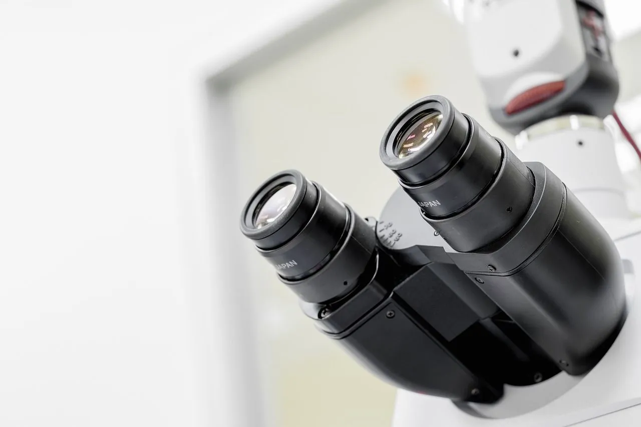
Illustration image Pixabay
Researchers face significant challenges when attempting to observe structures as small as the tubes forming the cytoskeleton of cells, with a diameter of only seven nanometers. Similarly, the synaptic cleft, which separates two nerve cells or a nerve cell and a muscle cell, measures between 10 and 50 nanometers. These dimensions are well below what traditional microscopes can capture, making these critical areas practically invisible to scientists. The lack of precise details at this scale limits the understanding of cellular mechanisms.
In light of these obstacles, researchers from the universities of Göttingen and Oxford, in collaboration with the University Medical Center Göttingen (UMG), have developed an innovative fluorescence microscope. This microscope relies on "single-molecule localization microscopy," which allows the visualization of structures with unprecedented precision. This method involves turning fluorescent molecules in a sample on and off to determine their exact position. Through this approach, it is possible to model the entire structure of the sample based on the precise localization of each fluorescent molecule.
The team led by Professor Jörg Enderlein at the Faculty of Physics of the University of Göttingen has introduced significant improvements to this technique. By integrating an ultra-sensitive detector and using specialized data analysis, they have managed to double the resolution, improving it from 10-20 nanometers to 5 nanometers. This advancement reveals previously invisible details, particularly in the junctions between two nerve cells, where the organization of proteins can now be observed with extreme precision.
In addition to its performance, this technology is notable for its ease of use and relatively low cost. The researchers emphasize that this new approach could be widely adopted in the research field, thanks in particular to the development of open-source software for data processing. This software, available to all, would allow other scientists to benefit from this advanced technology without major financial or technical obstacles.
The development of this high-resolution microscope represents a significant breakthrough for microscopy and cellular biology research. By making the invisible visible, it paves the way for new discoveries about the internal workings of cells and nano-scale interactions that were previously out of reach. This technology could thus transform our understanding of biological processes and offer new perspectives in the field of medical research.