Follow us on Google News (click on ☆)

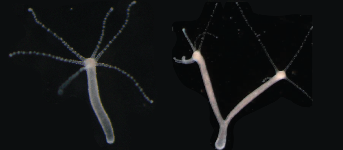
By applying, using an agar gel for four days, a slight pressure along the severed body of the hydra, scientists induced two topological defects, leading to the formation of two heads (left).
© Roux - UNIGE
This work reveals how external mechanical constraints can modify the symmetry points of the organism and influence its development. These symmetries, present in other more complex animal species, could play an important role in evolution. The study is published in the journal Science Advances.
In Greek mythology, the Hydra is a monster slain by Heracles, with each of its nine heads regrowing as soon as it is cut off. But the hydra is also a small aquatic creature from the jellyfish family, discovered in the 18th century, living in the calm freshwaters of lakes and ponds. Measuring only a few millimeters, it has a foot and a head equipped with fine tentacles. Its extraordinary regenerative abilities earned it the name of its mythological counterpart.
When a hydra is severed perpendicular to the body axis (between the foot and the head), it regenerates by forming a new head. By applying slight compression to the tissues regenerating the head, a team led by Aurélien Roux, full professor in the Department of Biochemistry of the Section of Chemistry and Biochemistry at the Faculty of Sciences of UNIGE, successfully induced the formation of viable two-headed hydras. A phenomenon that had never been observed before.
A two-headed hydra, produced by pressure on the severed body of the animal, capturing brine shrimp introduced into the environment to feed.
Thanks to a "topological defect"
"Inside the hydra, actin filaments are arranged parallel along the foot-head axis and converge at the head to form a 'topological defect' in the actin network," explains Yamini Ravichandran, a postdoctoral researcher in Aurélien Roux's team and first author of the study. "Our work shows that these topological defects in actin order play a central role in head regeneration, acting as mechanical organizers."
By simple pressure
By applying, using an agar gel for four days, a slight pressure along the severed body of the hydra, scientists induced two topological defects, leading to the formation of two heads. To verify if a defect creates a head, the researchers intended to remove these defects and observe if the animal could still regenerate. However, there is a difficult constraint to overcome, namely topology.
Parallel lines on a sphere always create two defects: the poles of the sphere. To eliminate them, pressure parallel to the actin filaments causes them to merge and disappear, forming a "donut"-shaped tissue, which cannot regenerate a head and eventually starves to death. The topology of the "donut" is unique, as it is the only one that can accept no defects and is absent in biology.
The hydra is a valuable study model due to its easily observable actin network, but the results obtained are applicable far beyond this species. Until now, how cells and tissues coordinate the forces that shape organisms is not well understood. The proposed concept is that genetics determine cell fate, which in turn dictates the forces that shape tissues.
This study shows that genetic factors and tissue mechanics are coupled—and thus act at the same level—to form a correct head in the hydra. As Aurélien Roux points out, "these results offer new insights into the mechanical signals that guide tissue repair and regeneration, with potential implications for understanding these processes in other organisms."
Fluorescence microscopy video showing the regeneration of a severed tissue into a two-headed hydra. The ordered muscle fibers are visible.
Fluorescence microscopy video showing the generation of a severed tissue into a ring that does not regenerate. The ordered muscle fibers are visible.